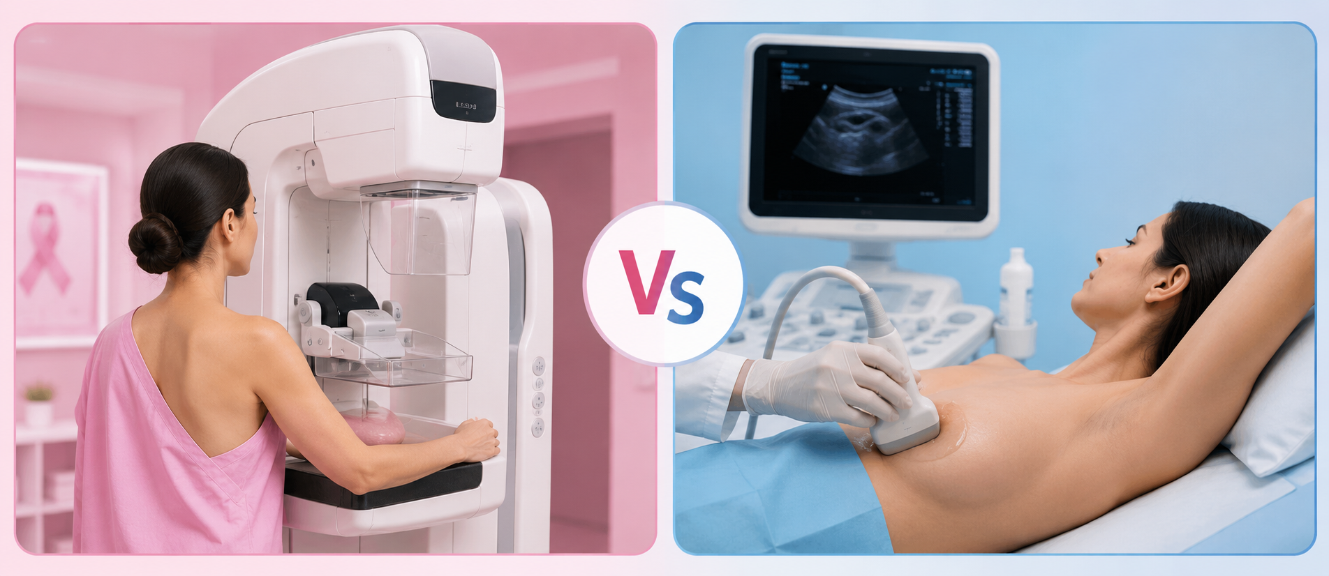

Once an oncologist recommends breast imaging, the next question is almost always the same, "Which test, and why?"
Two tests come up most often in breast health conversations: mammography and breast ultrasound. Both examine breast tissue and play a role in detecting cancer. But they work differently, serve different purposes and are suited to different clinical situations.
Comprehending what is a mammogram and breast ultrasound helps you walk into your appointment informed and less anxious about what to expect.
What Is a Mammogram and How Does It Work?
A mammogram is an X-ray picture of the breast. It uses low-dose radiation to produce detailed images of the breast tissue that enables radiologists to detect changes in the structure of the breast that cannot be felt in a physical exam. It remains the ideal choice for mammogram screening in women above the age of 40.
The Mammogram Procedure
During the mammogram procedure, each breast is placed on a flat platform and gently compressed between two plates. The compression may be slightly uncomfortable, but it is very important to spread the tissue out evenly, to lessen the amount of radiation needed, and to get sharper images. The entire process takes about 15 to 20 minutes. Two standard views are taken of each breast, one from the top and one from the side. The images are then reviewed by a radiologist trained in breast imaging.
Types of Mammograms
There are two primary types of mammograms used in clinical practice today.
2D digital mammography generates flat, 2D images of the breast. It is readily available as well as effective, especially for women with fatty breast tissue.
3D mammography, also called digital breast tomosynthesis, takes a series of images from multiple angles and reconstructs them into thin cross-sectional layers. This makes it considerably more useful for women with dense breast tissue, where overlapping tissue in a 2D image can obscure findings. It also reduces the number of call-backs for additional imaging because equivocal findings are easier to interpret.
Breast Cancer Mammogram: What It Can Detect
A breast cancer mammogram is most valuable for identifying microcalcifications. These are tiny calcium deposits that can be an early sign of malignancy. It also identifies masses, architectural distortions and asymmetry in breast tissue. These findings may appear on a mammogram years before a lump becomes palpable. This early detection window is precisely why routine mammogram screening matters.
What Is a Breast Ultrasound?
A breast ultrasound uses high-frequency sound waves rather than radiation to produce real-time images of breast tissue. It does not replace mammography, but it adds meaningful clinical information in specific situations.
Breast Ultrasound Procedure
The breast ultrasound procedure is straightforward and painless. A gel is applied to the breast skin, and a handheld transducer is moved across the surface. Sound waves pass through the tissue and bounce back to produce images on a monitor. Here, no compression is involved, and radiation exposure is almost zero. The scan typically takes 15 to 30 minutes depending on the area being evaluated.
When Is a Breast Ultrasound Used?
An ultrasound scan of the breast is particularly useful in the following scenarios-
1. Characterising a lump found on mammography or clinical examination
Ultrasound can determine whether a mass is solid or fluid-filled (a cyst). This distinction matters because simple cysts are almost always benign and require no further intervention.
2. Imaging dense breast tissue
Mammograms can miss lesions in women with dense breasts. It is because both the dense tissue and potential tumours appear white on X-ray. Ultrasound is not affected by tissue density and can reveal masses that a mammogram might obscure.
3. Guiding biopsy procedures
When a suspicious area needs tissue sampling, real-time ultrasound guidance ensures the needle is placed accurately.
4. Evaluating younger women
In women below 30, whose breast tissue tends to be dense, ultrasound is often the preferred first-line imaging tool.
Early-Stage Breast Cancer Ultrasound
A breast cancer ultrasound typically shows a malignant mass as an irregular, hypoechoic (darker than surrounding tissue) lesion with poorly defined margins. Early stage breast cancer ultrasound findings can include small solid nodules with suspicious characteristics, prompting further evaluation.
That said, ultrasound does not reliably detect microcalcifications. It is therefore most useful when used in conjunction with mammography, not as a substitute.
Mammography vs Breast Ultrasound: Choosing the Right Test
These two investigations are complementary, not competing. Mammography excels at screening and at detecting calcifications. Ultrasound excels at characterising masses and imaging dense tissue. In many clinical situations, both are used together to build a complete picture.
Your doctor's recommendation will depend on your age, breast density, symptoms, family history, and the specific question being answered. If a routine screening is what you need, mammography is the starting point. If a lump has already been found, or if your tissue is dense, an ultrasound is likely to follow.
A Note on Breast Ultrasound Price
The breast ultrasound price in India varies across facilities and cities, typically ranging from Rs. 800 to Rs. 3,000 depending on whether it is a focused or whole-breast scan. At top hospitals, both investigations are available with radiologist interpretation, and your treating doctor will advise which is appropriate for your situation.
Ending note
If you are residing in Bangalore, seeking guidance from a trusted breast cancer hospital in Bangalore or consulting experienced breast cancer doctors in Bangalore can help you understand which test is most appropriate for your situation. Neither test is better in an absolute sense. Each does something the other cannot. What matters is that the right test reaches the right patient at the right time.
If you are due for a mammogram screening, have noticed a change in your breast, or simply want clarity on where you stand, do not postpone that conversation with your doctor. Early investigation, even when nothing is found, gives you something valuable: certainty.
FAQs
Can a breast ultrasound detect cancer that a mammogram missed?
Yes, in some cases. Particularly in women with dense breast tissue, ultrasound scan breast can identify solid masses that appear invisible on a mammogram. The two investigations are most powerful when used together rather than in isolation.
At what age should I start mammogram screening?
Most guidelines recommend annual mammogram screening from age 40 onwards for women at average risk. Women with a family history of breast cancer or a known BRCA mutation may need to begin earlier, sometimes from age 30, and may require MRI in addition to mammography.
Is the mammogram procedure painful?
The compression involved in a mammogram causes temporary discomfort for most women, rarely significant pain. The sensation lasts only a few seconds per image. Scheduling your mammogram in the week after your menstrual period, when breast tissue is less tender, generally makes the experience more comfortable.